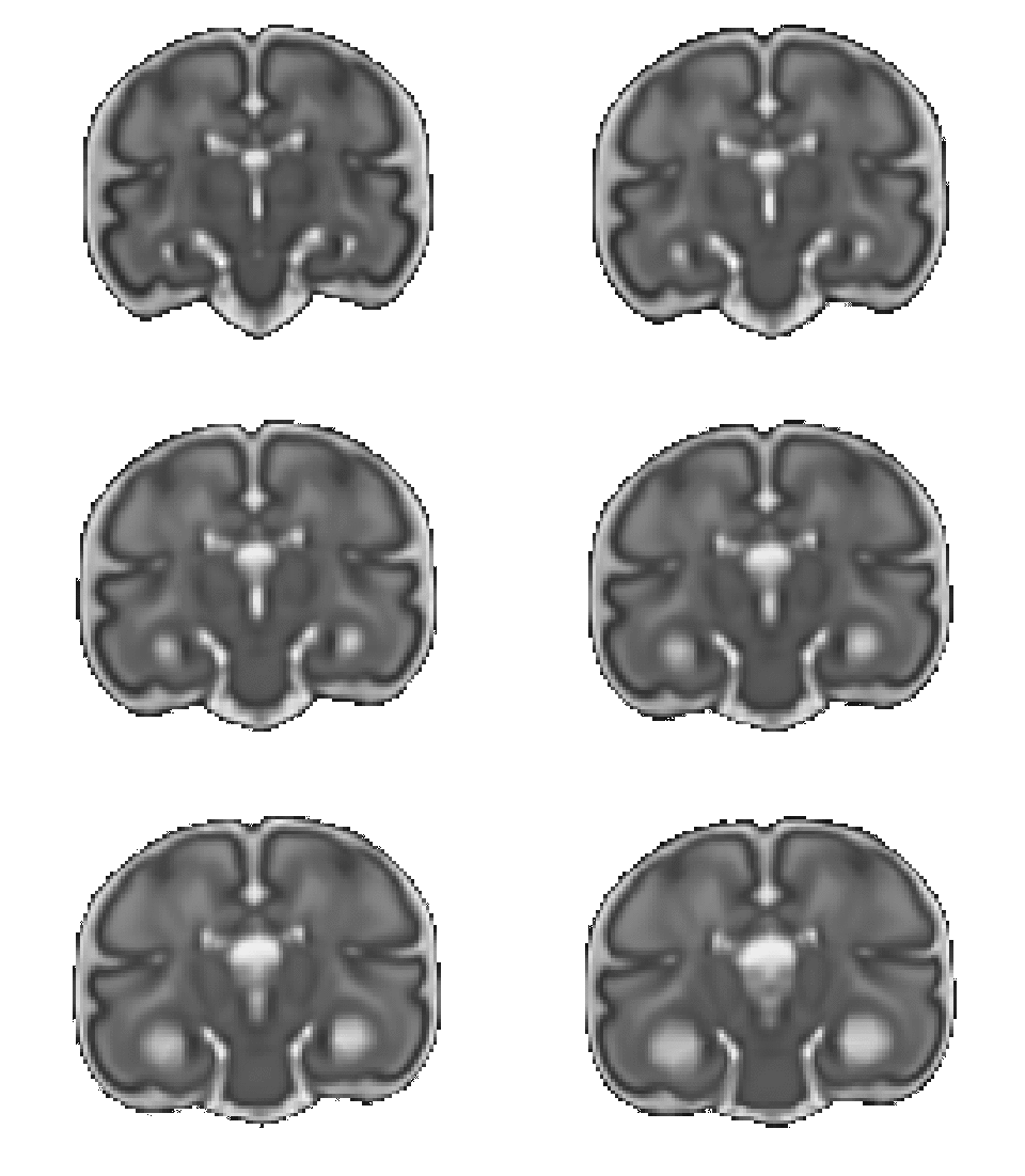
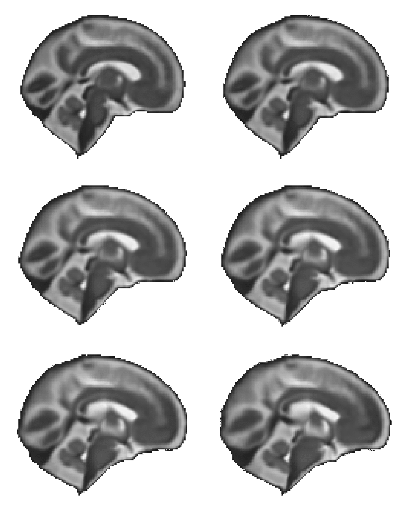
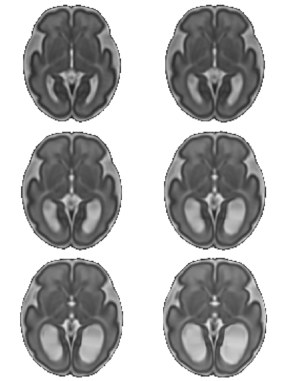
Below are displayed movies of the shape analysis tools described in our paper presented at the PIPPI workshop 2021.
Shape analysis pipeline for fetal brain MRI
This shape analysis pipeline is specifically tailored to the specificities of fetal MRI. Using diffeomorphic image registration, fetuses are compared to an age-machted template brain. Age differences between fetuses are corrected. Using PCA, a mean and variance analysis is performed on the subjects deformations to extract the modes of deformation that best characterize the anatomical variability of the dataset.
.gif)
Click here to display the reference trajectory of healthy brain growth during gestation
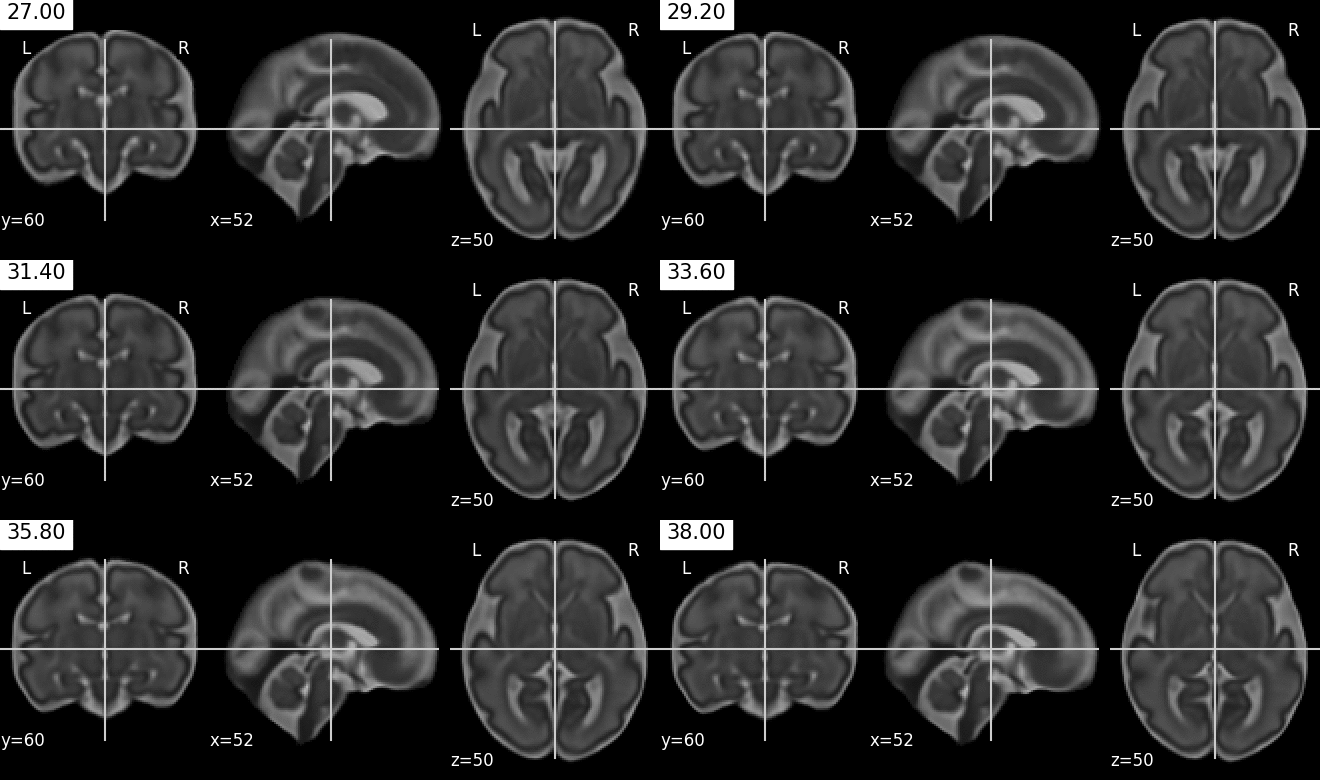
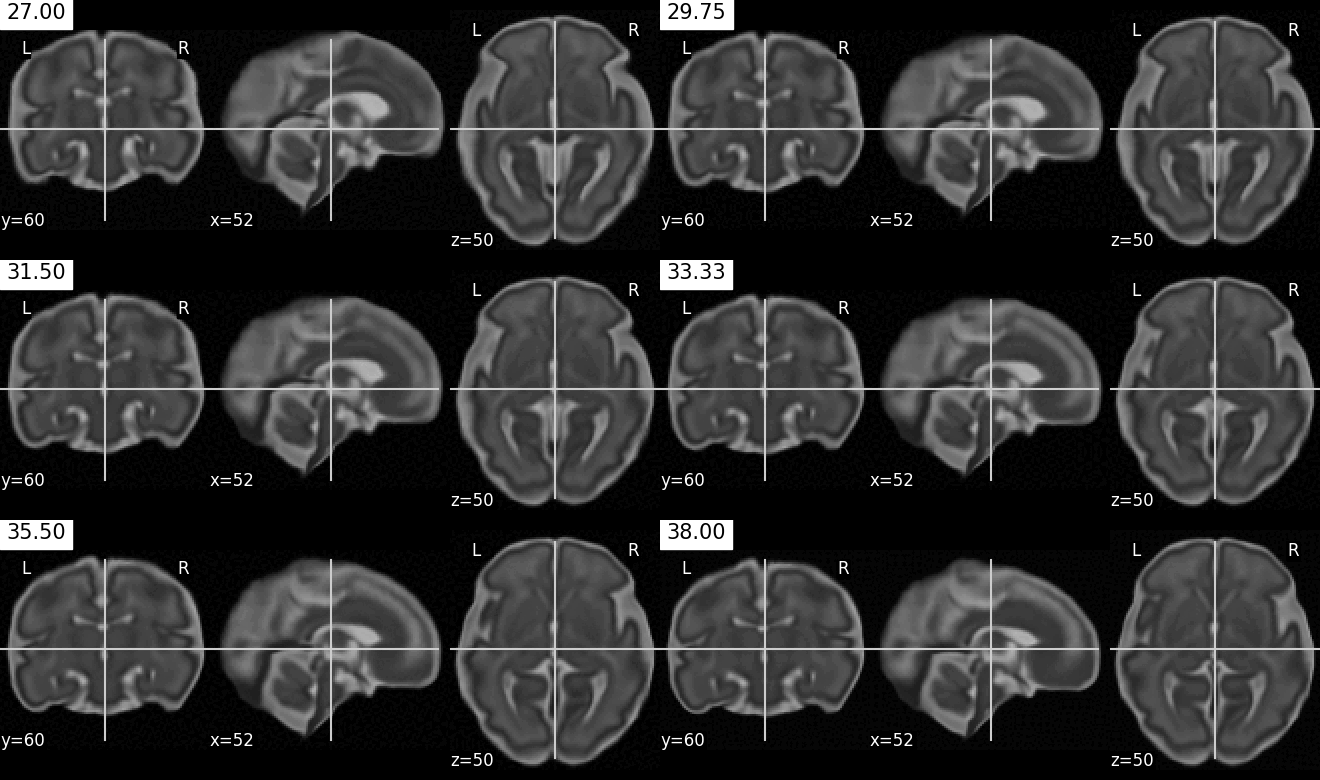
This is the geodesic trajectory of healthy brain growth (from 27 to 38 gestational weeks) used as reference to compute a distance from normality. Corresponds to the red curve.
Source of the healthy template brains used to compute the trajectory
Click here to display the transformation of the template brain towards Subject 2
Registration computes the transformation needed to map the healthy template at age 34 weeks towards the subject's brain, which has age 34 weeks (green arrow). The resulting transformation can be seen as a distance from normality.
.gif)
Click here to display the parallel transport of Subject 2 along the reference trajectory
To characterize a dataset of fetal brains (whether healthy or impaired), age differences between fetuses must be corrected: practically, this is done by transported the registration momenta to a common space. Parallel transport (blue arrows) transports the computed deformation to any time point along the red curve. Combined with geodesic shooting, we apply the reference growth dynamic to the subject's brain from 27 to 38 gestational weeks. The movie below illustrates how this brain, only observed at 34 weeks, would evolve during gestation (under the hypothesis that his growth rate is comparable to that of the healthy template).
Click here to display an example of deformation mode obtained on a dataset of fetuses with Corpus Callosum Agenesis
Second mode of deformation obtained by PCA on the initial vector fiels, between σ = 0 and σ = 4. This movie shows the deformation of an average healthy anatomy at 31 weeks of gestation towards the anatomy characterizing Corpus Callosum Agenesis.
.gif)
.gif)
.gif)
